
Pulse Portraits: Echocardiograms That Reveal the Real Story
An echocardiogram is more than a test. It is a portrait of rhythm, structure, and strength that helps guide confident decisions every day.
Heart health shapes the way people move, think, and enjoy everyday life, yet the heart rarely asks for attention until something feels slightly off. A flutter during a busy morning, a strange pressure after climbing a flight of stairs, or a wave of fatigue at the wrong moment can spark concern long before a diagnosis appears. An echocardiogram steps in as the quiet storyteller of the heart, revealing details that only sound waves can uncover. Local imaging professionals use advanced technology to capture moving pictures of the heart, turning those subtle symptoms into meaningful information. Patients walk away with answers, clinicians gain clarity, and the entire community gains stronger support for wellness. An echocardiogram is more than a test. It is a portrait of rhythm, structure, and strength that helps guide confident decisions every day.
The Purpose Behind Detailed Heart Imaging
A heart accomplishes an incredible amount of work from morning to night, making its performance one of the most important parts of personal health. An echocardiogram captures the heart in motion with real-time ultrasound images that show how the chambers contract, how the valves operate, and how blood flows throughout the body. Patients often feel reassured when they see how smoothly everything functions or relieved when they finally understand the cause behind their symptoms. The test offers the kind of visibility that cannot be achieved through a stethoscope alone.
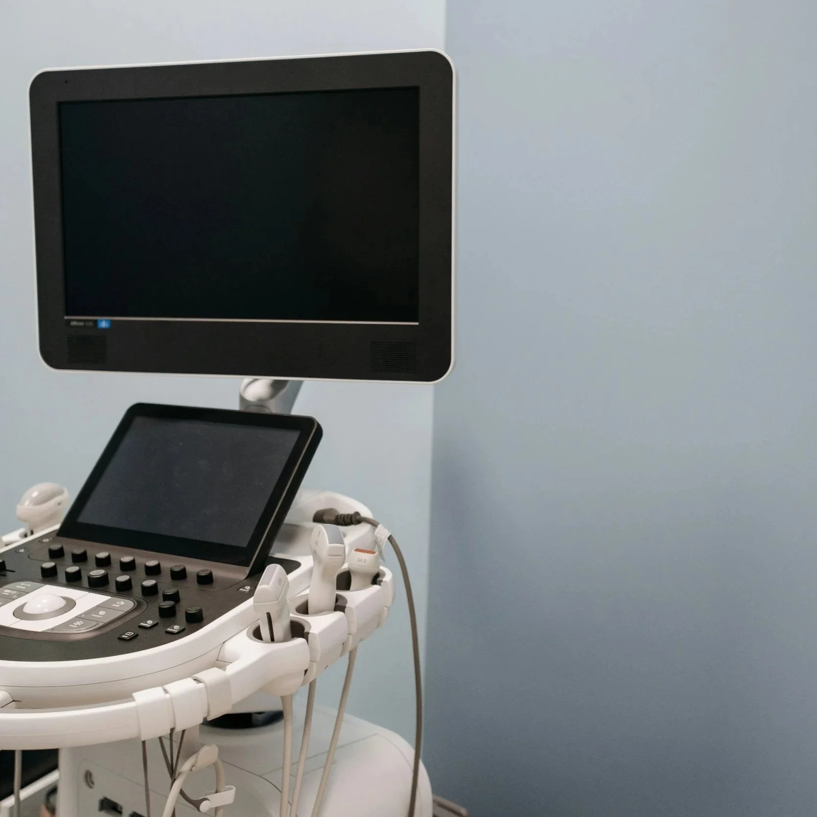
Imaging specialists use multiple angles to create a full picture of cardiac performance. A small handheld transducer glides across the chest while sound waves reflect off various heart structures. Those reflections appear on screen as moving images, giving clinicians a clear look at the mechanics behind every single beat. Patients appreciate the test's gentle, noninvasive nature and the immediate sense of understanding it provides. With each view, the story of the heart unfolds in greater detail, offering a level of clarity that supports accurate and timely decisions.
Why Noninvasive Imaging Creates A Better Experience
Echocardiograms offer a surprisingly peaceful experience compared to many medical tests. Patients lie comfortably while the sonographer applies warm gel and guides the transducer over the chest. There are no needles, no radiation, and no recovery period. The entire appointment feels far less intimidating than many expect, especially once they learn they can breathe normally throughout the process. Anxiety often fades as soon as the first images appear.
A noninvasive test also means patients of all ages can participate without added risk. Children, pregnant individuals, older adults, and those with chronic health conditions can undergo the procedure without concern. Local imaging teams focus on maintaining a relaxed atmosphere, answering questions as they go, and helping patients feel supported through every step. That comfortable experience encourages people to seek evaluation sooner rather than later, strengthening overall cardiac wellness in the community.
How Blood Flow Patterns Tell A Deeper Story
Blood flow reveals essential details that help clinicians understand how well the heart works under different conditions. Doppler technology, often included in echocardiogram studies, uses color and waveform displays to show the speed and direction of blood movement. When blood flows smoothly, the images reflect predictable patterns. When flow becomes uneven or turbulent, the images highlight areas where the heart may be compensating for valve problems or pressure imbalances.
Clinicians rely on these images to identify circulatory concerns that may not be apparent on a physical exam. Changes in pressure, slight leaks, or irregular movement can alert healthcare teams to early warning signs before major symptoms develop. Patients experiencing shortness of breath, swelling, or unexpected fatigue often find meaningful answers through these blood flow assessments. By visualizing the heart's internal motions, clinicians can recommend treatment paths that support healthier long-term outcomes.
Why Valve Performance Plays Such A Key Role
Heart valves act as carefully timed gates that direct blood flow with precision. When a valve does not open wide enough or does not close completely, the heart must work harder to compensate. Echocardiograms capture these subtle changes clearly, allowing clinicians to identify concerns early. Patients who hear terms like regurgitation or stenosis often feel overwhelmed at first, but seeing the valve in motion helps make the diagnosis easier to understand.
Structural issues within valves can progress slowly, and early detection helps prevent strain on the heart. Local imaging specialists focus on capturing crisp, informative views that help cardiologists develop targeted treatment plans. Whether a patient requires medication adjustments or ongoing monitoring, the insights from valve imaging help guide every decision. Community-based cardiac imaging centers provide accessible care, making it easier for patients to maintain follow-up appointments.
How Wall Structure Reflects Heart Strength
The heart's muscular walls adapt based on the body's needs, but certain conditions place additional stress on those muscles. High blood pressure, chronic illness, or genetic factors may cause the walls to thicken over time. An echocardiogram helps clinicians evaluate whether those changes affect the heart's ability to pump effectively.
Imaging reveals how the walls move with each beat and whether any areas struggle to contract or relax. Patients often find comfort in seeing how the heart responds to medication or lifestyle adjustments over time. Local imaging teams make routine monitoring simple, helping clinicians track changes and tailor treatment plans. When patients understand the structural strengths and stress points within their hearts, they play a more active role in their long-term care.
The Community Benefit Of Accessible Echo Imaging
Local access to echocardiogram services helps communities address cardiac concerns quickly and effectively. People do not need to travel far or wait weeks for a study, which encourages earlier evaluation. Early testing helps prevent major emergencies and supports faster recovery when symptoms arise.
Accessibility also strengthens the relationship between healthcare providers and their patients. With quick access to imaging results, clinicians can respond with timely recommendations that help maintain stability. Local imaging professionals often partner closely with primary care providers and specialists to streamline communication. That partnership enhances community wellness and builds trust in the local healthcare system.
How Early Imaging Supports Prevention
Cardiac conditions often develop quietly, with subtle signs that may not draw attention right away. An echocardiogram helps reveal early patterns that could evolve into more serious problems. When the heart begins working harder or circulation becomes inefficient, these early signs appear in the images. Patients who receive early intervention often avoid major complications later on.
Preventive cardiac care creates healthier communities overall. People feel empowered to take charge of their heart health when they receive clear explanations and reliable imaging. Local imaging centers help make these preventive steps part of routine wellness rather than emergency response. A simple scan today can prevent a crisis tomorrow, allowing families to continue their routines with greater peace of mind.
What Stress Echo Testing Adds To The Picture
Stress echocardiograms add another layer of insight by showing how the heart responds to increased demand. Clinicians capture images before and after exercise or medication that raises the heart rate. Patients dealing with exertion-related discomfort or unexplained fatigue often benefit from the broader perspective that stress imaging provides.
These studies help identify issues that only appear under physical pressure. When the heart struggles to meet increased demand, images reveal areas with reduced blood flow or weakened muscle segments. Local imaging teams support patients throughout the process, helping them stay calm and comfortable as they move through each stage. The information gained helps clinicians develop accurate guidance that addresses symptoms and protects long-term health.
Monitoring Chronic Conditions With Echo Imaging
Chronic illnesses frequently influence cardiac function over time. Conditions like hypertension, diabetes, kidney disease, and autoimmune disorders can gradually affect how efficiently the heart works. Regular echocardiograms create a visual record of these changes, helping clinicians understand how each condition shapes overall heart performance.
Patients often find the images reassuring because they offer concrete evidence of progress or areas that need attention. Consistent monitoring helps guide treatment adjustments and allows for timely intervention when necessary. Local imaging availability makes it easier for patients to maintain follow-up care, strengthening the partnership between clinicians and the community.
Building Confidence Through Clear Imaging
Patients gain confidence when they receive clear, understandable results from their echocardiogram. The visual nature of the images helps demystify complex medical information, allowing people to learn more about their heart without feeling overwhelmed. Many patients express relief after their study because they finally understand what their body has been trying to communicate.
Clear imaging also strengthens communication between clinicians and patients. When explanations align with visual evidence, people feel more engaged and empowered in their care plans. Local imaging teams contribute to that confidence by guiding patients through each step of the process in a calm, friendly manner.
The Value Of Professional Skill And Technique
Quality echocardiograms depend heavily on the skill of the imaging professional. Sonographers use their training to capture the best angles, adjust to different body types, and identify subtle variations in movement. Their expertise transforms a simple test into a powerful diagnostic tool.
Local imaging centers invest in ongoing education and up-to-date technology to maintain accuracy. Patients benefit from consistent, reliable imaging that supports informed decision-making. High-quality equipment and experienced professionals help deliver results that clinicians trust and patients appreciate.
How Positive Experiences Encourage Long-Term Care
A comfortable experience during an echocardiogram encourages patients to return for follow-up care. Calm communication, warm gel, and a supportive environment reduce anxiety and make the appointment feel approachable. Patients who enjoy a pleasant experience often become more proactive about monitoring their heart health.
Positive experiences also strengthen relationships between imaging centers and the community. People who feel valued and supported are far more likely to recommend services to family members, friends, and neighbors. That trust helps enhance the overall heart health of the community.
How Routine Echo Imaging Supports Community Wellness
Routine echocardiograms contribute to a healthier community by helping residents stay informed, proactive, and engaged in their cardiac care. When people have access to clear imaging and reliable guidance, they feel better equipped to make decisions that protect long-term health.
Echo imaging supports early detection, timely intervention, and ongoing stability across neighborhoods. The combination of professional skill, advanced technology, and warm care creates a foundation for strong community wellness. The heart is one of the most important parts of life, and an echocardiogram offers the insight needed to keep it functioning at its best.
Act Now: Essential Echocardiogram FAQs You Should Know
What symptoms often lead to an echocardiogram?
Common symptoms include chest discomfort, irregular heartbeat, shortness of breath, swelling, or unexplained fatigue.
Is an echocardiogram safe for all ages?
Echocardiograms use sound waves and are safe for children, adults, and older individuals.
How long does the test usually last?
Most echocardiograms take between 30 and 60 minutes, depending on the views needed.
Do I need special preparation before the appointment?
Standard echocardiograms require no special preparation, though stress echo tests may involve instructions based on the type of stress used.
How often should follow-up imaging be scheduled?
Follow-up frequency depends on individual health needs and medical history. Clinicians guide ongoing monitoring based on each patient's condition.
Cardiovascular Specialty Imaging offers various on-site ultrasound services and mobile Ultrasound Services to physicians, imaging centers, hospitals, and nursing facilities throughout Northwest Florida. Our services include abdominal ultrasound, renal ultrasound, arterial ultrasound, venous evaluation, and echocardiogram. Call us today.